Lipid Droplets Embedded in a Model Cell Membrane Create a Phospholipid Diffusion Barrier
Abstract
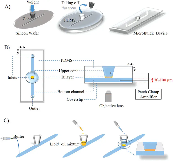
Abstract Lipid droplets (LDs) are ubiquitous, cytoplasmic fat storage organelles that originate from the endoplasmic reticulum (ER) membrane. They are composed of a core of neutral lipids surrounded by a phospholipid monolayer. Proteins embedded into this monolayer membrane adopt a monotopic topology and are crucial for regulated lipid storage and consumption. A key question is, which collective properties of protein-intrinsic and lipid-mediated features determine spatio-temporal protein partitioning between phospholipid bilayer and LD monolayer membranes. To address this question, a freestanding phospholipid bilayer with physiological lipidic composition is produced using microfluidics and micrometer-sized LDs are dispersed around the bilayer that spontaneously insert into the bilayer. Using confocal microscopy, the 3D geometry of the reconstituted LDs is determined with high spatial resolution. The micrometer-sized bilayer-embedded LDs present a characteristic lens shape that obeys predictions from equilibrium wetting theory. Fluorescence recovery after photobleaching measurements reveals the existence of a phospholipid diffusion barrier at the monolayer–bilayer interface. Coarse-grained molecular dynamics simulation reveals lipid specific density distributions along the pore rim, which may rationalize the diffusion barrier. The lipid diffusion barrier between the LD covering monolayer and the bilayer may be a key phenomenon influencing protein partitioning between the ER membrane and LDs in living cells.