Quantifying the influence of the ion cloud on SAXS profiles of charged proteins

Abstract
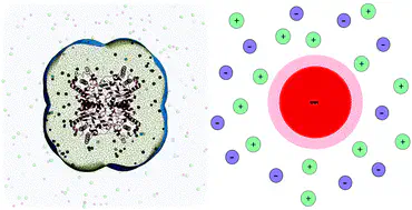
Small-angle X-ray scattering (SAXS) is a popular experimental technique used to obtain structural information on biomolecules in solution. SAXS is sensitive to the overall electron density contrast between the biomolecule and the buffer, including contrast contributions from the hydration layer and the ion cloud. This property may be used advantageously to probe the properties of the ion cloud around charged biomolecules. However, in turn, contributions from the hydration layer and ion cloud may complicate the interpretation of the data, because these contributions must be modelled during structure validation and refinement. In this work, we quantified the influence of the ion cloud on SAXS curves of two charged proteins, bovine serum albumin (BSA) and glucose isomerase (GI), solvated in five different alkali chloride buffers of 100 mM or 500 mM concentrations. We compared three computational methods of varying physical detail, for deriving the ion cloud effect on the radius of gyration $R_g$ of the proteins, namely (i) atomistic molecular dynamics simulations in conjunction with explicit-solvent SAXS calculations, (ii) non-linear Poisson–Boltzmann calculations, and (iii) a simple spherical model in conjunction with linearized Poisson–Boltzmann theory. The calculations for BSA are validated against experimental data. We find favorable agreement among the three computational methods and the experiment, suggesting that the influence of the ion cloud on $R_g$, as detected by SAXS, may be predicted with nearly analytic calculations. Our analysis further suggests that the ion cloud effect on $R_g$ is dominated by the long-range distribution of the ions around the proteins, as described by Debye–Hückel theory, whereas the local salt structure near the protein surface plays a minor role.