Viral fusion proteins of classes II and III recognize and reorganize complex biological membranes
Abstract
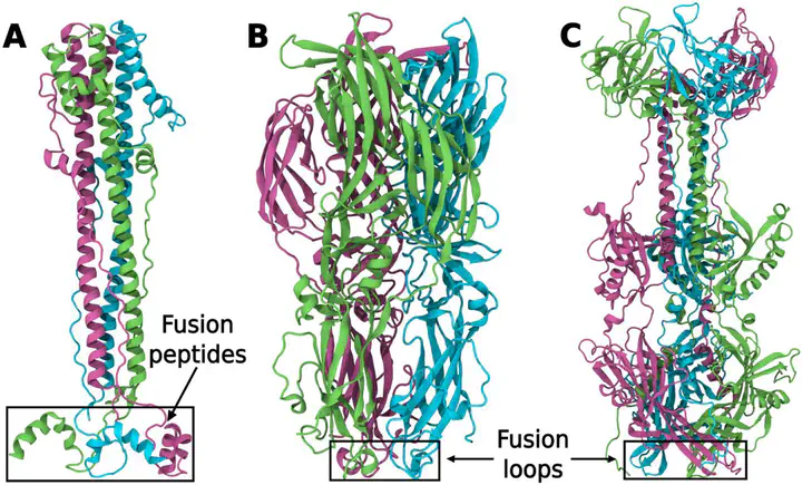
Viral infection requires stable binding of viral fusion proteins to host membranes, which contain hundreds of lipid species. The mechanisms by which fusion proteins utilize specific host lipids to drive virus–host membrane fusion remains elusive. We conducted molecular simulations of classes I, II, and III fusion proteins interacting with membranes of diverse lipid compositions. Free energy calculations reveal that class I fusion proteins generally exhibit stronger membrane binding compared to classes II and III — a trend consistent across 74 fusion proteins from 13 viral families as suggested by sequence analysis. Class II fusion proteins utilize a lipid binding pocket formed by fusion protein monomers, stabilizing the initial binding of monomers to the host membrane prior to assembling into fusogenic trimers. In contrast, class III fusion proteins form a lipid binding pocket at the monomer–monomer interface through a unique fusion loop crossover. The distinct lipid binding modes correlate with the differing maturation pathways of classes II and III proteins. Binding affinity was predominantly controlled by cholesterol and gangliosides as well as via local enrichment of polyunsaturated lipids, thereby locally enhancing membrane disorder. Our study reveals energetics and atomic details underlying lipid recognition and reorganization by different viral fusion protein classes, offering insights into their specialized membrane fusion pathways.